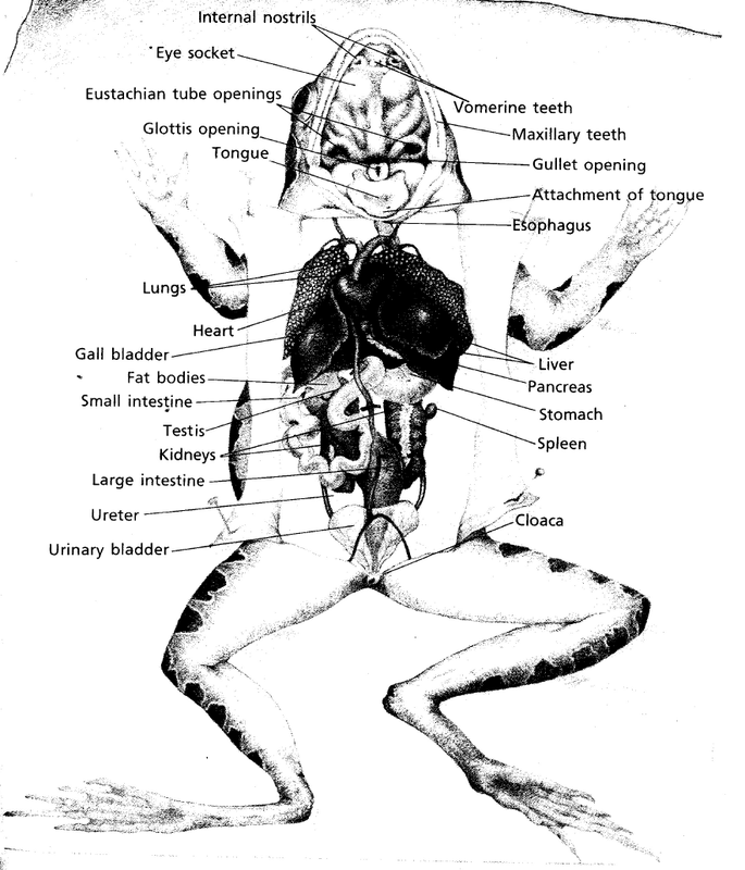
Secondly, locate the two prominent vomerine teeth located behind the mid-region of the upper jaw. Firstly, locate the fine maxillary teeth lining the upper jaw.

Locate the glottis (leads to the lungs) and oesophagus opening (leads to the stomach).Carefully cut the jaw joints on each side of the mouth to enable you to open the mouth wide.Reposition the frog to lie on its dorsal side.The cloacal opening provides the function of exit for all urinary, reproductive, and digestive systems. Identify the cloaca, located at the specimen’s posterior end.As the frog is deceased, this will appear as a cloudy eyelid attached at the bottom of the eye however, it would appear clear in a living frog. Look closely at the eyes and attempt to find the frog’s third eyelid this is the nictitating membrane that moistens and protects the eye.You will find these on the back side of the eyes. Find the round tympanic membranes that form the frog’s external sound receptors.These function as a means of respiration. Looking at the head, identify the 2 external nares at the head’s tip.The frog’s hind limbs are divided into a thigh, lower leg, and foot. You will see that each forelimb includes an upper arm, forearm, and hand. The frog uses 4 limbs to travel and move, making it a tetrapod. Observe the frog appendages that have evolved to adapt to terrestrial life.Place the preserved frog on the dissecting tray with the dorsal surface facing up.Provide each workstation with the following materials:.This should be done in a well-ventilated room and appropriate PPE should be worn. To prepare frog specimens, open the sealed bag with scissors, pour out any excess liquid and rinse the specimen with tap water.PREPARATION - BY LAB TECHNICIAN General Preparations This practical requires only a few materials and takes a short time, yet provides students from a range of year levels with engaging, memorable lesson in anatomy. Exploring the internal systems of frogs provides a tangible example of various anatomical structures and how they function. It provides insight into evolutionary adaptations, such as the transition from an aquatic life to life on land. Dissection of preserved frogs is an established introductory activity into vertebrate anatomy and mature body systems. They provide a great model organism for a range of biological studies including, evolution, behaviour, anatomy, and physiology. In animals, the transport of materials within the internal environment for exchange with cells is facilitated by the structure and function of the circulatory system at cell and tissue levels (for example, the structure and function of capillaries) (ACSBL058)įrogs are vertebrates with specialised amphibian characteristics and behaviours.In animals, the exchange of nutrients and wastes between the internal and external environments of the organism is facilitated by the structure and function of the cells and tissues of the digestive system (for example, villi structure and function), and the excretory system (for example, nephron structure and function) (ACSBL057).In animals, the exchange of gases between the internal and external environments of the organism is facilitated by the structure and function of the respiratory system at cell and tissue levels (ACSBL056).

FROG DISSECTION DIAGRAM LABELED PDF
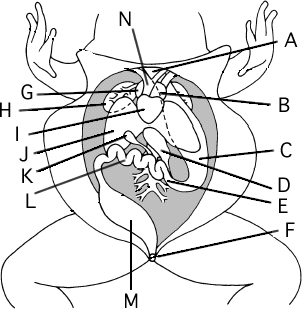
Frog Dissection: Complete Guide – includes external anatomy, mouth, and the organs of the abdominal cavity, download available in pdf and google doc.


 0 kommentar(er)
